Fig. 10
Download original image
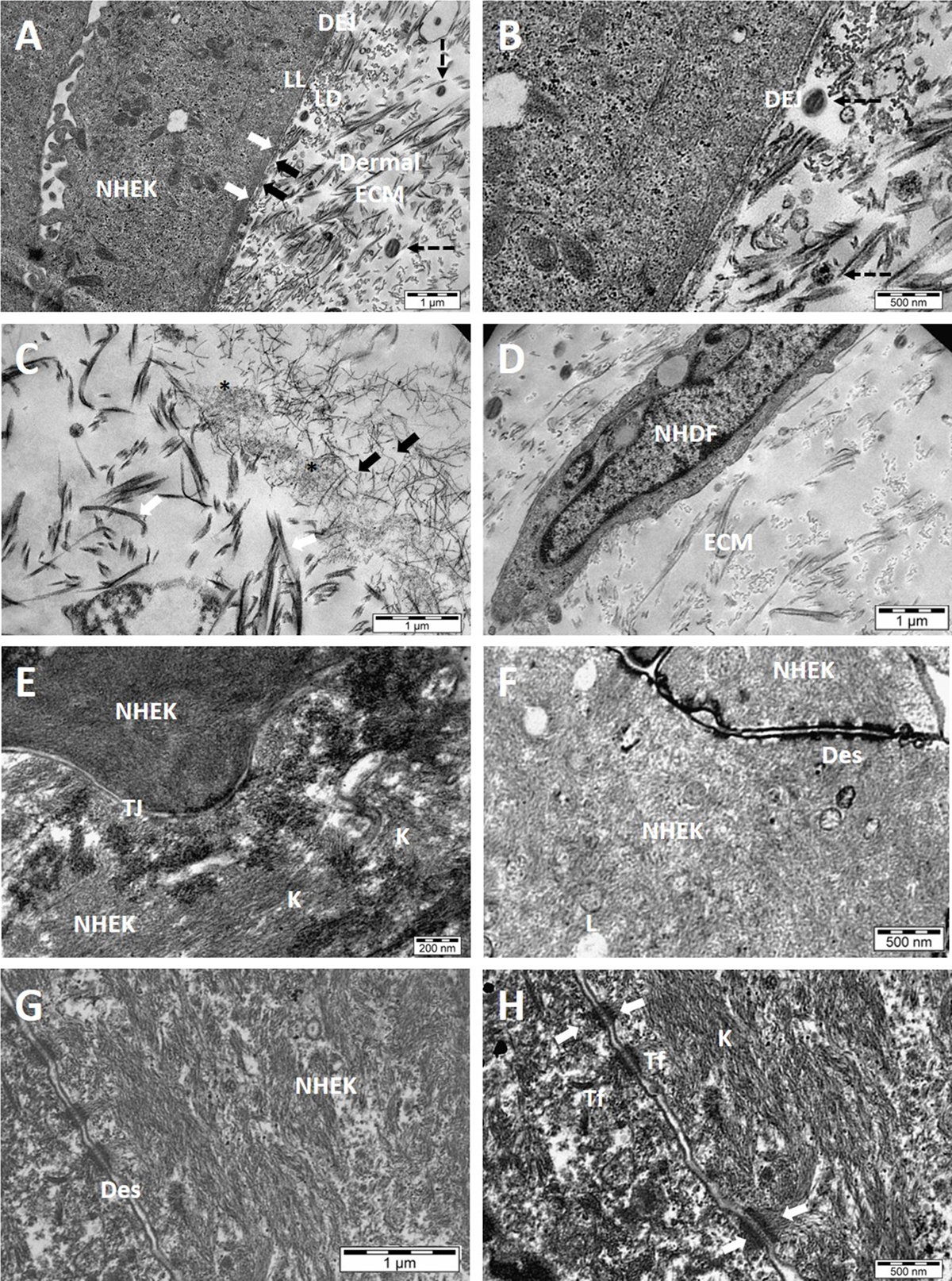
Ultrastructure analysis of HSE. TEM images (A, B) revealed dermal-epidermal junction (DEJ) that separates epidermis from the dermis through basement membrane consisting of lamina lucida (LL) and lamina densa (LD). LD was connected with the collagen matrix by loops of anchoring fibrils (black arrows). The hemidesmosomes (white arrows) were connected with tonofibrils (Tf) in keratinocytes. Collagen fibers were shown as black dotted arrows. TEM images (C, D) demonstrated collagen fibrils (Col. I) with their characteristic banded pattern (white arrows). Microfibrils or fibrillins (black arrows) were also secreted by fibroblasts, forming a scaffold that was detected near collagen fibrils. Amorphous or “immature” elastin was secreted and aggregated (asterisks) on microfibrils. Collagen fibers (black dotted arrow) appeared when collagen fibrils combined to form fibers. Normal Human Dermal Fibroblasts (NHDF) were also seen as embedded in ECM. In TEM images (E, F, G, H) epidermis showed tight junction (TJ) between the keratinocytes. Keratinocytes showed abundant intracellular filament of keratins (K) and upper epidermal layers showed lipid droplets (L). Tonofibrils (Tf) in keratinocytes (NHEK) were made of tonofilaments (keratin intermediate filaments), that appear converging to form spot like connections or desmosomes (Des or white arrows); N = 2, n = 3.