Open Access
Fig. 6
Download original image
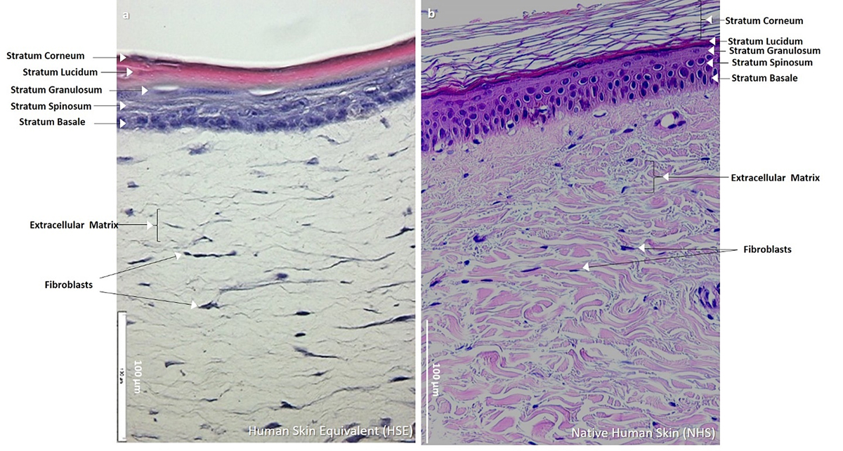
(a) HSC as HSE. H&E-stained histological image of HSC obtained from 3D-CC-III (cf. Fig. 5C′′) was able to best recapitulate epidermal differentiation, morphogenesis and organization similarly as found in (b) NHS (origin: human leg; histology: different staining protocol) and thus named as HSE. HSE showed two structurally distinct layers of skin the outer epidermal layer, and the underlying thicker dermal layer. The epidermal part showed well differentiated layers of keratinocytes namely stratum basale, spinosum, granulosum, and corneum. The staining differences between HSE and NHS are due to a different H&E protocol used to stain NHS at the Institute of Pathology, Ruhr University Bochum, Germany, where the NHS tissue was obtained.