Open Access
Figure 1
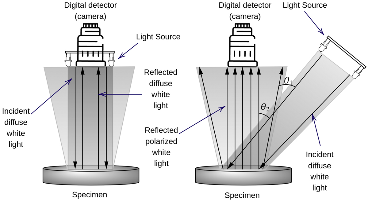
Download original image
Schematic representation of conventional imaging spectroscopy using diffuse light (left) and special imaging spectroscopy using polarized light (right). The relative positions of light sources for the two imaging spectroscopies are indicated. Reflected diffuse white light arises from the Light source (6 light-emitting diodes arranged in the circle) perpendicular to the sample and reflected polarized light (right) arises from the light source positioned at Brewster’s angle (for biological tissues, approximately 530 degrees). The degree of light polarization is 95.4%, while angular diffusion of the light source is ± 1:6 (the difference between the angles θ1 and θ2), which through convolution spectra gives Opto-magnetic Imaging Spectroscopy.